Home/
Unlabelled
/Bone Cross Section Diagram Labeled / Long Bone High Res Stock Images Shutterstock / Bone is found in the shafts of long bone and consists of various cylindrical units named as haversian system 47.
Bone Cross Section Diagram Labeled / Long Bone High Res Stock Images Shutterstock / Bone is found in the shafts of long bone and consists of various cylindrical units named as haversian system 47.
Bone Cross Section Diagram Labeled / Long Bone High Res Stock Images Shutterstock / Bone is found in the shafts of long bone and consists of various cylindrical units named as haversian system 47.. Diagram with articular cartilage, marrow, spongy bone, medullary cavity, endosteum, diaphysis, and periosteum. can be used for personal and commercial purposes. Bone tissue cross section diagram human oasissolutions co. Schematic diagram of compact and spongy bones. Spinal cord crosssection images stock photos vectors shutterstock. This is a cross section through decalcified bone.
Fibrous layer (with fibroblasts) cellular layer ( chondroblasts). Diagram with articular cartilage, marrow, spongy bone, medullary cavity, endosteum, diaphysis, and periosteum. In a cross section of a bone we can see two types of bone tissue: Cross section diagram skin cross section labeled body part chart removable wall graphic. Cross section of the forearm through the flexor carpi ulnaris muscle:
1 Schematic Drawing Of A Longitudinal Section Through A Long Bone Download Scientific Diagram from www.researchgate.net Diagram with articular cartilage, marrow, spongy bone, medullary cavity, endosteum, diaphysis, and periosteum. In a cross section of a bone we can see two types of bone tissue: Labeled anatomical structure and location. Bone tissue cross section diagram human oasissolutions co. From wikimedia commons, the free media repository. See labeled cross sections of the human body now at kenhub. The line will be indicated by an actual line, or with positions labelled with letters on. Bone cross section vector illustration diagram.
The line will be indicated by an actual line, or with positions labelled with letters on.
Skin anatomy diagram description illustration skin stock. Internal structure of the dicotyledonous stem by openstax. Cross section of the forearm through the flexor carpi ulnaris muscle: Explaned distal and proximal epiphysis. From wikimedia commons, the free media repository.
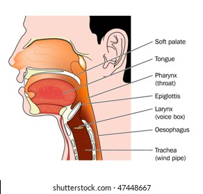
Nose Mouth Throat Cross Section Labeled Stock Vector Royalty Free 47448667 from image.shutterstock.com Skin anatomy diagram description illustration skin stock. Compact bone is the outer layer and the spongy bone forms the inner layer. Each system contains haversian canals surrounded by concentric lamellae of bone tissue 48. Related posts of bone cross section labeled. Explaned distal and proximal epiphysis. Labeled diagram with brain sections. These pictures of this page are about:cross section of ground compact. Vector illustration bone cell types diagram.
Bone is found in the shafts of long bone and consists of various cylindrical units named as haversian system 47.
For example, to read this diagram literally, since the cartilage can be seen. Medically reviewed by the healthline medical network — written by the healthline editorial team — updated on january 20, 2018. From wikimedia commons, the free media repository. This is a cross section through decalcified bone. In a cross section of a bone we can see two types of bone tissue: Cross section spinal cord stock vector royalty free 74226298. Bone tissue cross section diagram human oasissolutions co. Diagram with articular cartilage, marrow, spongy bone, medullary cavity, endosteum, diaphysis, and periosteum. Bone cross section vector illustration diagram. Cross section of bone diagram. There are trabeculae in spongy bone which gives its sponge like appearance. Diagram of channel cross section leaf cross section diagram label worksheets. I am not an expert on this subject, so i was wondering if anyone could put their input on i don't like way you've shown the cartilage.
Bone marrow is the soft, highly vascular and flexible connective tissue within bone cavities. Jump to navigation jump to search. Each system contains haversian canals surrounded by concentric lamellae of bone tissue 48. Fibrous layer (with fibroblasts) cellular layer ( chondroblasts). As shown in figure 2.
These pictures of this page are about:cross section of ground compact. In a cross section of a bone we can see two types of bone tissue: See labeled cross sections of the human body now at kenhub. Cross section diagram skin cross section labeled body part chart removable wall graphic. Labeled diagram with brain sections. Explaned distal and proximal epiphysis. There are trabeculae in spongy bone which gives its sponge like appearance. Vector illustration scheme of bone cross section. Compact bone is the outer layer and the spongy bone forms the inner layer. From wikimedia commons, the free media repository. Cross section diagram diagram spinal cord cross section wiring diagram review. Schematic diagram of compact and spongy bones. 2048 x 3713 jpeg 341 кб.
Cross section spinal cord stock vector royalty free 74226298 bone cross section. Diagram with articular cartilage, marrow, spongy bone, medullary cavity, endosteum, diaphysis, and periosteum. can be used for personal and commercial purposes.
Bone Cross Section Diagram Labeled / Long Bone High Res Stock Images Shutterstock / Bone is found in the shafts of long bone and consists of various cylindrical units named as haversian system 47.
Reviewed by MAXenzy
on
Maret 31, 2021
Rating: 5


 Reviewed by MAXenzy
on
Maret 31, 2021
Rating:
Reviewed by MAXenzy
on
Maret 31, 2021
Rating:


Post a Comment